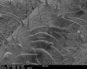
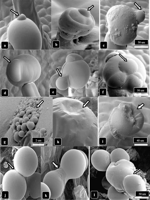
Scanning Eletron Microscopy photograph of the leaf surface of Solanum
By A Mystery Man Writer
Last updated 26 May 2024

Download scientific diagram | Scanning Eletron Microscopy photograph of the leaf surface of Solanum granuloso-leprosum Dunal. A – Unicelular tector trichomes; B – tector trichome, note that there are projections at the trichome base; C – tector trichome, note that there is a larger projection/ramification at the trichome base; D – tector trichome, note that there are two larger projection/ramification at the trichome base; E – tector trichome, note that there are three larger projection/ramification at the trichome base; F – tector trichome, note that there are four larger projection/ramification at the trichome base; G – tector trichome, note that there are five larger projection/ramification at the trichome base; H – tector trichome, note that there are six larger projection/ramification at the trichome base; I – tector trichome, note that there are eight larger projection/ramification at the trichome base; J – another angle from the six ramification tector trichome; and K – multicelular and multisseriated tector trichome, note the thick secondary cell wall. Scale Bars = 20 μm. from publication: Anatomy, histochemistry and micromorphology of leaves of Solanum granuloso-leprosum Dunal | In the present work the anatomical, histochemical and micromorphological features of S. granuloso-leprosum leaves were approached in order to evaluate its characteristics associated with its pioneer role. Glandular and non-glandular trichomes were observed on both epidermal | Micromorphology, Solanum and Plant Anatomy | ResearchGate, the professional network for scientists.

Leaf epidermal characters of Brazilian species of Solanum section Torva as taxonomic evidence

Vibrant Leaf Surface

Topography-Driven Shape, Spread, and Retention of Leaf Surface Water Impacts Microbial Dispersion and Activity in the Phyllosphere
Scanning electron microscopic (SEM) images captured at 60×

Genetic Control of Glandular Trichome Development: Trends in Plant Science

Scanning Eletron Microscopy photograph of the leaf surface of Solanum
Microorganisms, Free Full-Text

Morphological characterisation of trichomes

Scanning electron microscopic images of the lower leaf epidermis
Scanning Electron Microscope Collection of Photo Prints and Gifts #3

Scanning electron microscopy images of A. dauci on the leaf surface of
Recommended for you
-
 Microscopic photograph of C. sativa leaf-trichomes; CST26 May 2024
 -
 Cannabis trichomes: what are they?26 May 2024
 -
![Trichomes under the microscope [OC] : r/trees](https://external-preview.redd.it/pnBO78R5Pw74_zu9JV3UOedoj9snOp2heo01imkXZxo.jpg?width=640&crop=smart&auto=webp&s=d2155f112398f0d4fa6557ade4b21c460f1dcc49) Trichomes under the microscope [OC] : r/trees26 May 2024
Trichomes under the microscope [OC] : r/trees26 May 2024 -
 Trichomes - Development, Types, Functions and Microscopy Science photos, Macro and micro, Scanning electron micrograph26 May 2024
 -
 Glandular trichome development, morphology, and maturation are influenced by plant age and genotype in high THC-containing cannabis (Cannabis sativa L.) inflorescences, Journal of Cannabis Research26 May 2024
 -
 File:Trichomes under a microscope.jpg - Wikimedia Commons26 May 2024
 -
 Trichome Microscope - Kosmic Kitchen26 May 2024
 -
![PDF] Plant trichomes and the biomechanics of defense in various systems, with Solanaceae as a model](https://d3i71xaburhd42.cloudfront.net/178c62764e9bfe37678388eac4b5ec3f1e5eb034/33-Figure1-1.png) PDF] Plant trichomes and the biomechanics of defense in various systems, with Solanaceae as a model26 May 2024
PDF] Plant trichomes and the biomechanics of defense in various systems, with Solanaceae as a model26 May 2024 -
 Plant Leaf Trichome (hibiscus Sp.) Photograph by Dennis Kunkel Microscopy/science Photo Library - Pixels26 May 2024
 -
Click sound on and take a look at the wonders of cannabis under a26 May 2024

You may also like
-
 25PCS Tattoo Transfer Paper Stencil Carbon Thermal Tracing Hectograph Sheets26 May 2024
 -
 84pcs Silicone Beads Kit,9 Colors Silicone Beads for Keychain Making,15mm Round,14mm Polygonal Silicone Beads,16mm Wooden Beads,with 2m Lanyard,626 May 2024
 -
 Fraggle Rock' Movie Gets a Pair of Writers (Exclusive) – The26 May 2024
 -
 Denim Blue Puffy Short Eyelash Yarn 68178 Ice 100 Gram, 180 Yards Micro Fiber26 May 2024
 -
 Whirlpool 5-cu ft High Efficiency Stackable Steam Cycle Front-Load Washer (Chrome Shadow) ENERGY STAR26 May 2024
 -
 Fiebing's Pro Leather Dye 4 oz. Oxblood - Hill Leather Company26 May 2024
 -
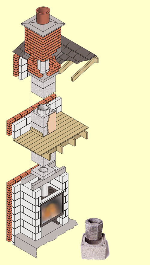 dm pumice chimney26 May 2024
 -
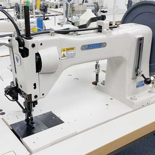 THOR GA 733 Extra Heavy Duty Walking Foot Sewing Machine for Extremely Heavy Materials and Thread - Sunny Sewing Center26 May 2024
 -
 Hydro Flask 32 oz All Around Travel Tumbler Trillium26 May 2024
 -
 Rosineer 10x7 Parchment Paper, Non Stick, Pre-Cut26 May 2024